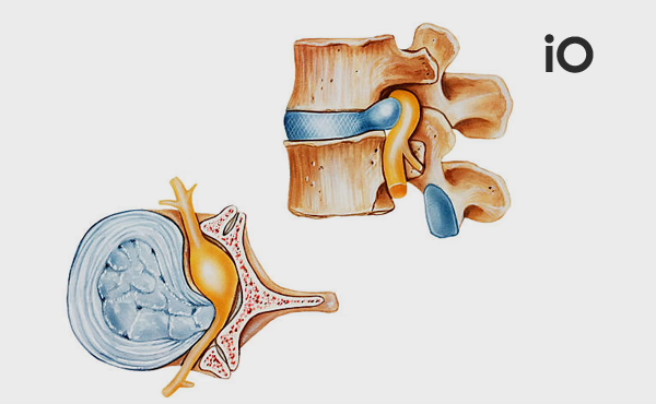
When surgical intervention is required for a herniated disc, the choice of surgical method varies depending on the surgeon's expertise, the level of the herniation, its location within the spinal canal, and factors such as calcification (ossification).
Here are the details of the commonly used microscopic and endoscopic methods in herniated disc surgery:
Microscopic Herniated Disc Surgery
Microscopic surgery is regarded as the gold standard in spinal surgery and is one of the most commonly used techniques. Its features include:
- Enhanced Visualisation: Performed under a microscope, this surgery allows the surgeon to view the nerve root, spinal cord, and other structures in a magnified and detailed manner.
- Small Incisions: The surgery is performed through a skin incision of approximately 3-5 cm.
- Targeted Intervention: The approach is made from the side where the herniation is located. Muscles are gently retracted without damaging the spine's anatomical integrity. A small piece of bone from the posterior lamina is removed (laminectomy).
- Controlled Disc Removal: After carefully separating the ligamentous tissue over the spinal cord, the herniated disc material is removed.

Endoscopic Herniated Disc Surgery
Endoscopic surgery is a minimally invasive technique performed using a camera-equipped imaging system. Its advantages and types include:
- Minimal Tissue Damage: This technique involves small incisions and is one of the least invasive options.
- Two Technique Options:
- Transforaminal Endoscopic Approach: This approach provides access to herniations located outside or at the edges of the spinal canal. It minimises tissue and bone damage but carries a slightly higher risk of nerve trauma due to proximity to the nerve root.
- Interlaminar Endoscopic Approach: Similar to the microscopic technique, this method is ideal for central herniations and involves entering through a comparable incision site.
Which Surgical Technique Should Be Preferred?
The choice of surgical technique depends on the location of the herniation within the spine, the degree of canal narrowing, and calcification. Examples include:
- Foraminal and Far Lateral Herniations: The transforaminal approach is more suitable.
- Centrally Located Herniations: The microscopic or interlaminar endoscopic method is advantageous.
- Pelvic Bone Height Considerations: At the L5-S1 level, the transforaminal method may be challenging. In such cases, the microscopic or interlaminar technique is preferable.
Advantages and Disadvantages
- Endoscopic Methods: These offer faster recovery in the early stages and result in less post-operative pain due to minimal tissue damage. However, dura (spinal cord membrane) repair is more challenging compared to the microscopic method, and conversion to open surgery may be required in some cases.
- Microscopic Methods: These provide a wider field of view and make repair more manageable in the event of complications.
Long-Term Outcomes
Regardless of the method used, long-term outcomes are generally similar. Therefore, determining the most appropriate surgical technique for each patient is crucial.
Conclusion
In herniated disc surgery, the choice of method should be tailored to the patient's condition and the characteristics of the herniation. Factors such as the level of the herniation, its location, calcification, and nerve root anomalies must be thoroughly evaluated. Before surgery, patients should be informed of the advantages and disadvantages of all techniques, and the most suitable surgical option should be presented.
Remember: The process can be managed more safely and effectively with an experienced spinal surgeon.

